Dr Aditya Malik FRCSEd

Home  /  Gallery

Imaging & Surgery

Case Gallery

A small selection of imaging illustrating the kind of work described elsewhere on this site. Every image is anonymised, and nothing here identifies a patient.

Shoulder & Elbow

Upper limb

The problem on the left, the result on the right. Radiographs from shoulder and elbow reconstruction, replacement and fracture surgery.

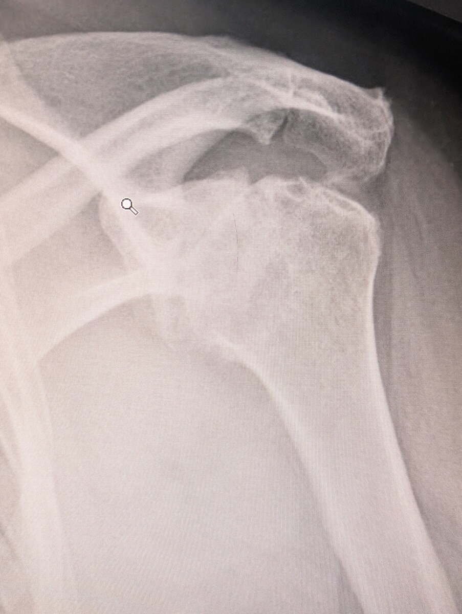
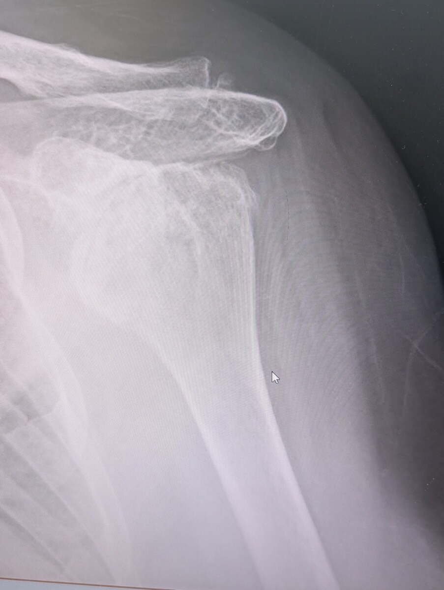
Before
Radiograph of rotator cuff arthropathy, the humeral head risen against the acromion
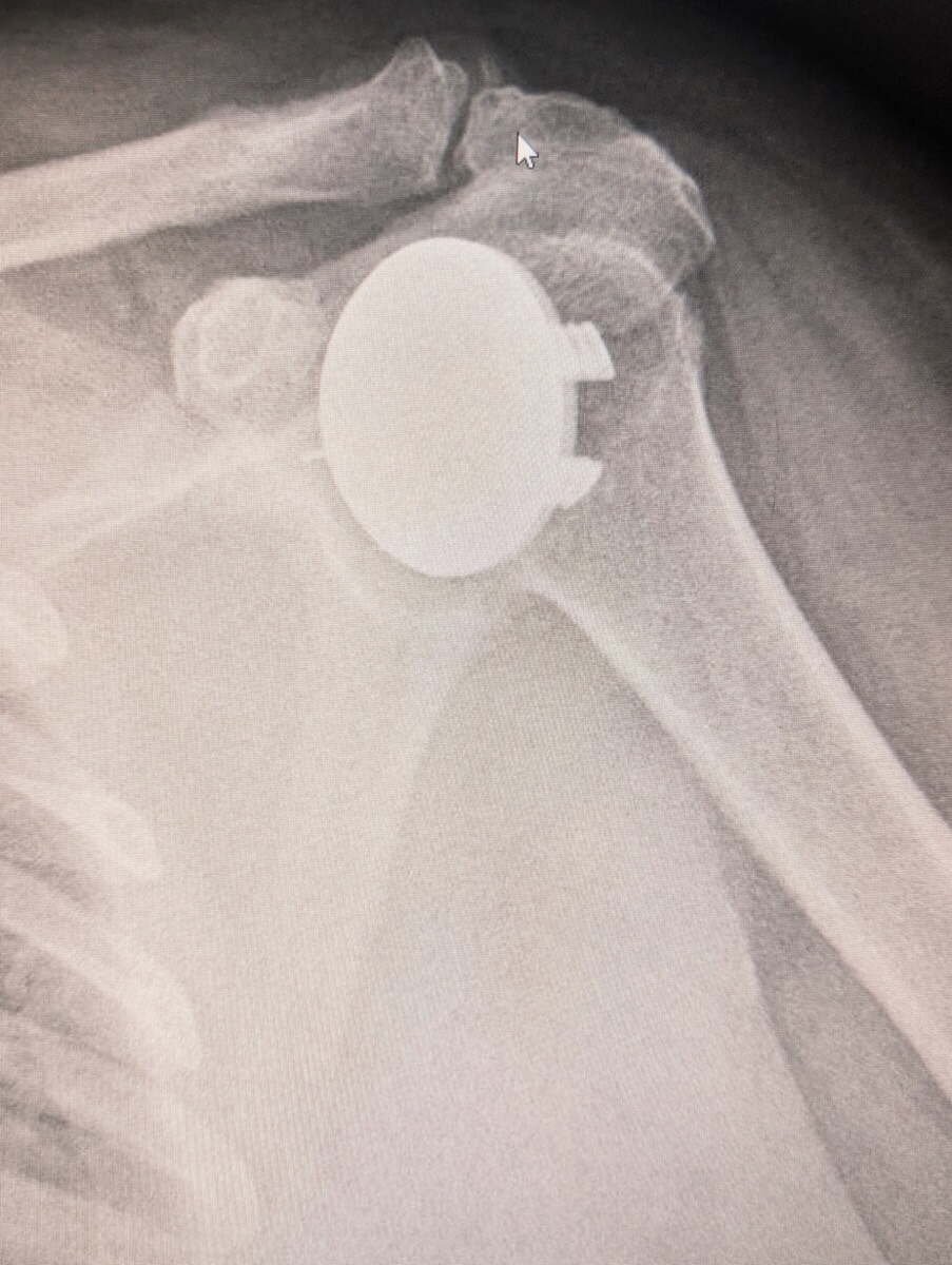
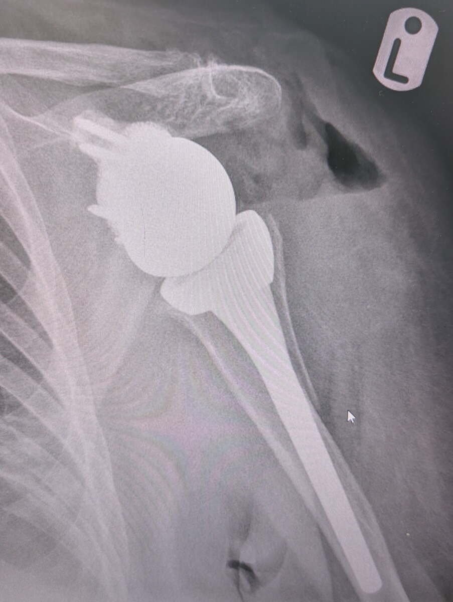
After
Radiograph of a reverse total shoulder replacement for cuff arthropathy
Reverse Shoulder Replacement

Rotator cuff arthropathy. The cuff has failed and the humeral head has risen against the acromion. A reverse replacement lets the deltoid do the work the cuff no longer can.

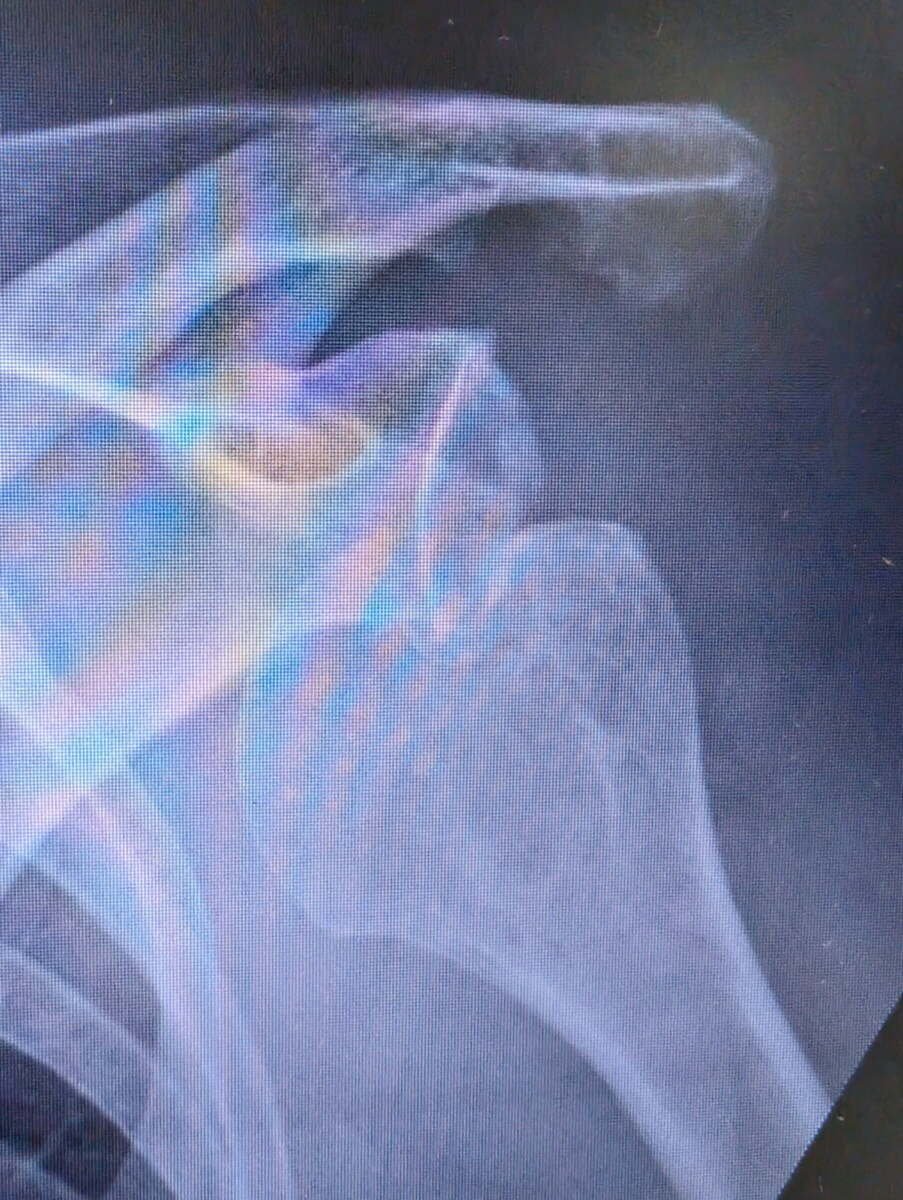
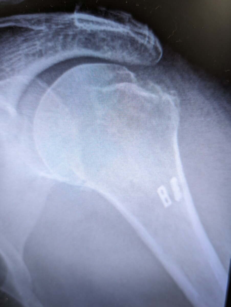
Before
Radiograph of a shoulder with severe glenohumeral osteoarthritis before surgery
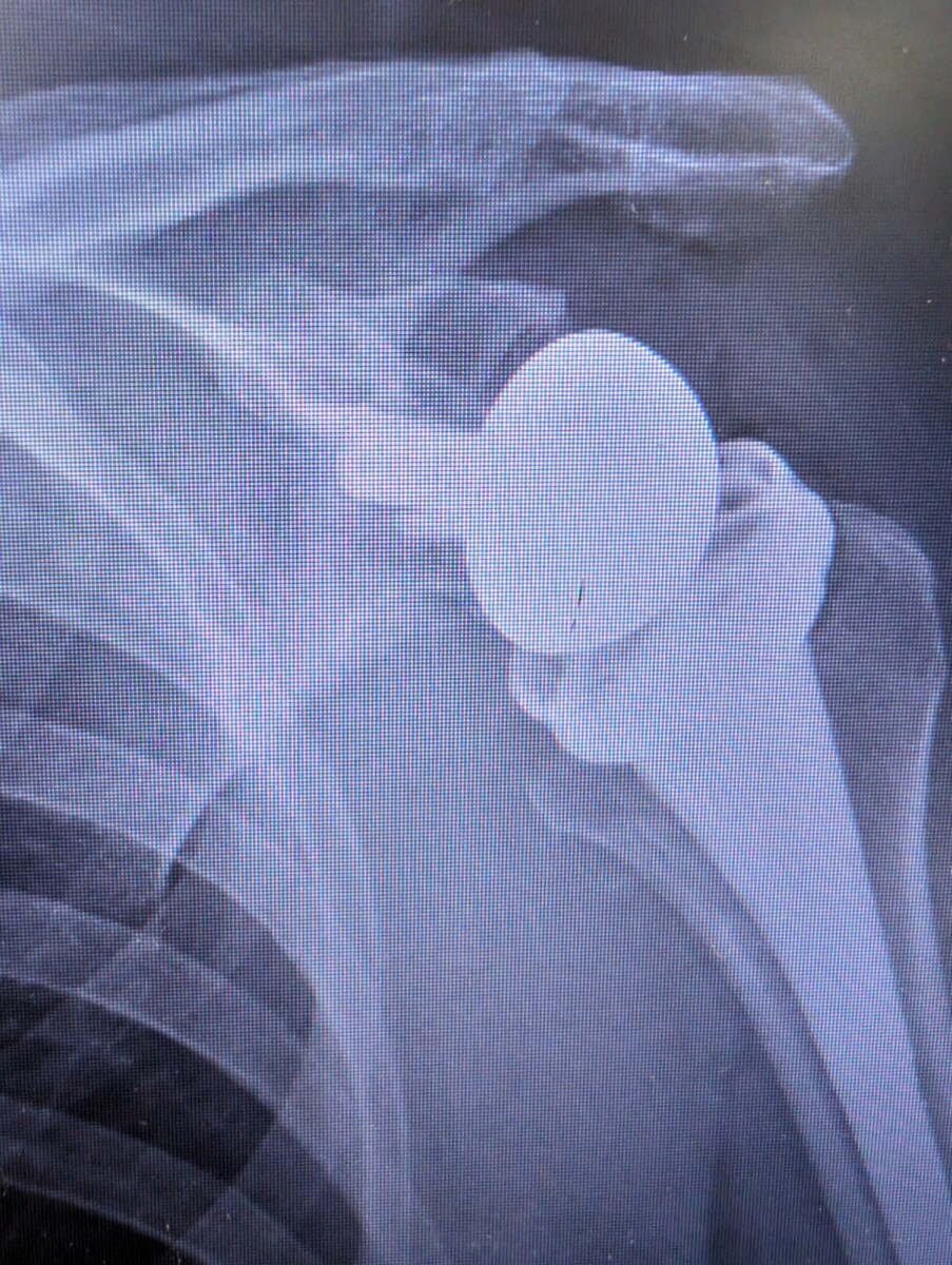
After
Radiograph showing an anatomic stemless total shoulder replacement
Anatomic Shoulder Replacement

Severe glenohumeral arthritis with the joint space worn away. A stemless anatomic replacement recreates the normal ball and socket, and depends on an intact rotator cuff.

Before
Radiograph of a chronic locked anterior shoulder dislocation
After
Radiograph of a reverse total shoulder replacement for chronic locked dislocation
Reverse Replacement for Chronic Dislocation

A shoulder locked out of joint for months in an elderly patient. Putting it back would not have held, so the joint was reconstructed.

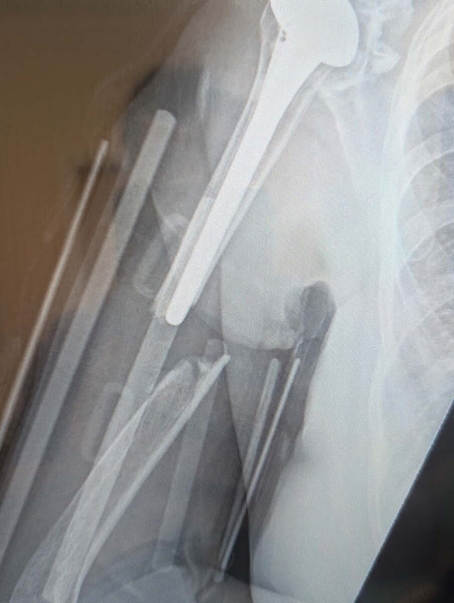
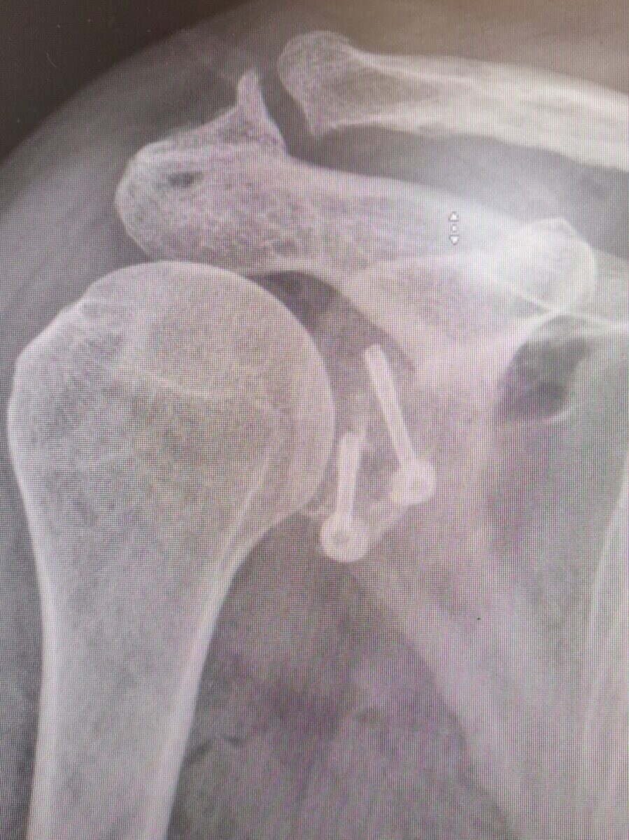
Before
Radiograph of a periprosthetic humerus fracture around a shoulder replacement stem
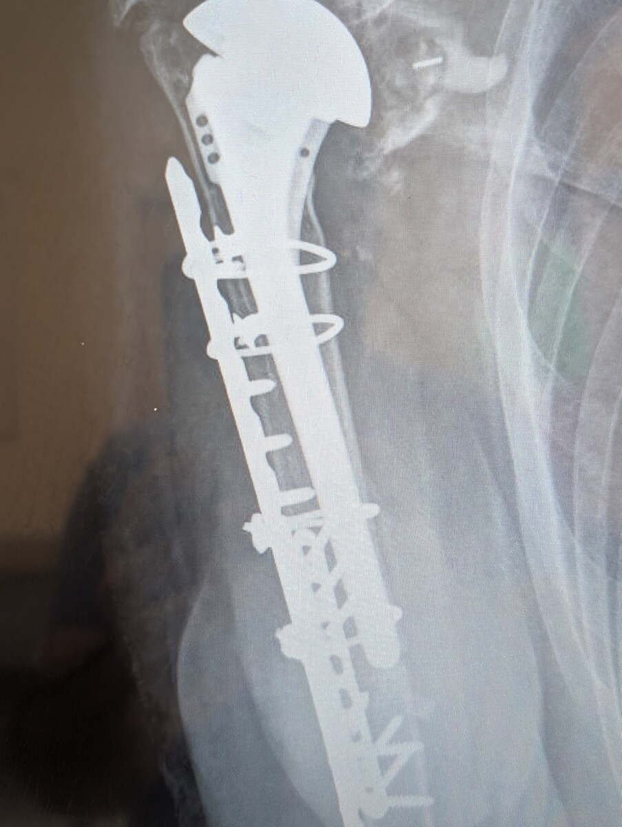
After
Radiograph showing a periprosthetic humerus fracture fixed with two plates, the original implant preserved
Fracture Around a Shoulder Replacement

A humerus broken around an existing replacement stem. Fixed with two plates and cables, keeping the original implant rather than revising the whole reconstruction.

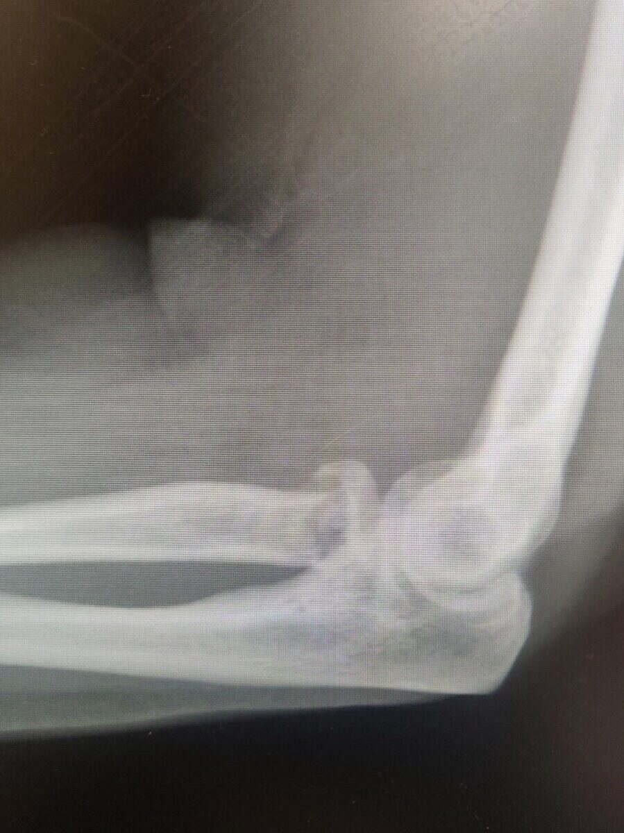
Before
Radiograph of a radial head fracture of the elbow
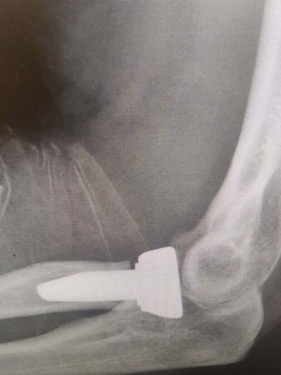
After
Radiograph of an uncemented radial head replacement
Radial Head Replacement

A radial head fracture too comminuted to repair reliably, treated with an uncemented replacement.

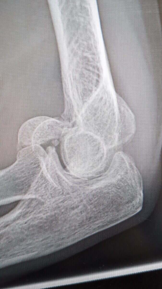
Before
Radiograph of a capitellum shear fracture of the elbow
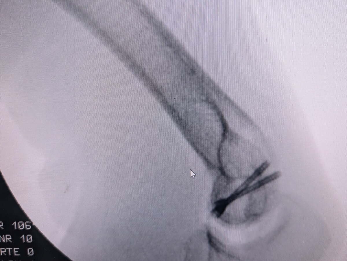
After
Radiograph of a capitellum fracture fixed with headless compression screws
Capitellum Shear Fracture

A shear fracture of the capitellum fixed with two headless compression screws, buried beneath the cartilage so nothing sits proud of the joint surface.

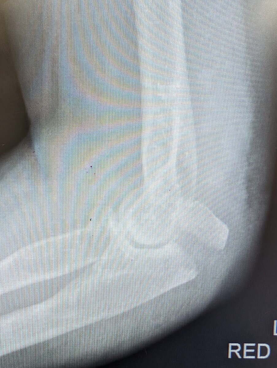
Before
Radiograph of an olecranon fracture of the elbow
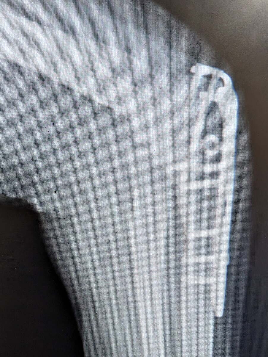
After
Radiograph of an olecranon fracture fixed with a locking plate
Olecranon Fracture

The point of the elbow, fixed with a locking plate.

Radiograph of a shoulder after a Latarjet procedure for instability
Latarjet Procedure

For recurrent instability where bone has been lost from the socket. The coracoid is transferred to rebuild the glenoid and acts as a sling across the front of the joint.

Radiograph of a repaired greater tuberosity fracture using suture anchors and endobuttons
Greater Tuberosity Repair

A fractured greater tuberosity held with suture anchors and endobuttons, restoring the cuff attachment without bulky metalwork.

Radiograph of a distal biceps tendon repaired to the radial tuberosity with an endobutton
Distal Biceps Repair

The biceps tendon reattached to the radial tuberosity with an endobutton, through a small incision at the front of the elbow.

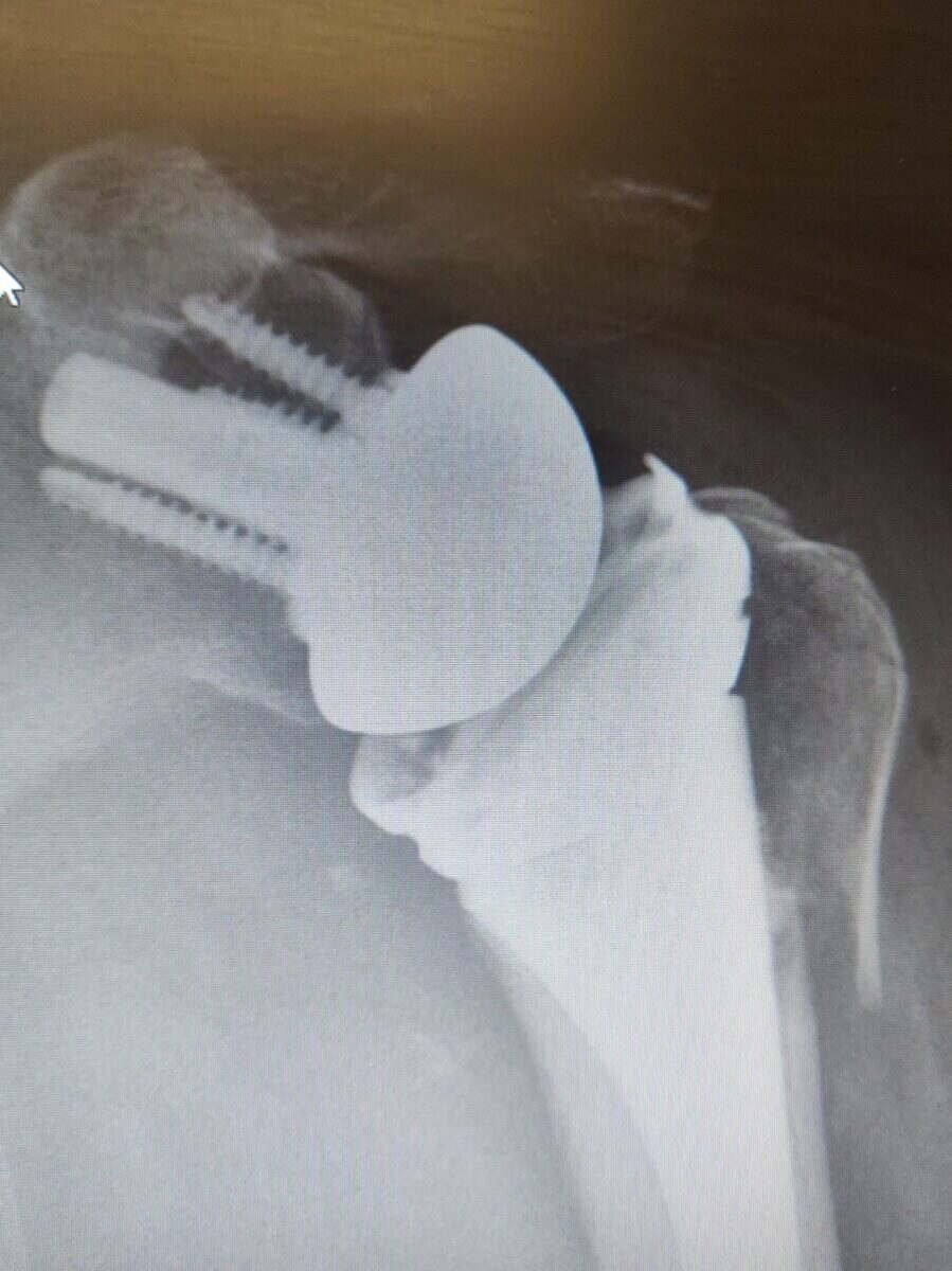
Radiograph of a left reverse total shoulder replacement for an unreconstructible fracture
Reverse Replacement after Fracture

A proximal humerus fracture too comminuted to reconstruct reliably, treated with a reverse replacement.

MRI of the shoulder in the ABER sequence showing a Bankart lesion
Bankart Lesion on MRI

An MRI taken in the ABER position, showing the labrum torn from the front of the socket after dislocation.

Hip & Knee

Lower limb

Before and after. Joint replacement is judged on alignment, on restoration of leg length and offset, and on how reliably the plan was carried out. Occasionally it is judged on how a failure is put right.

Before
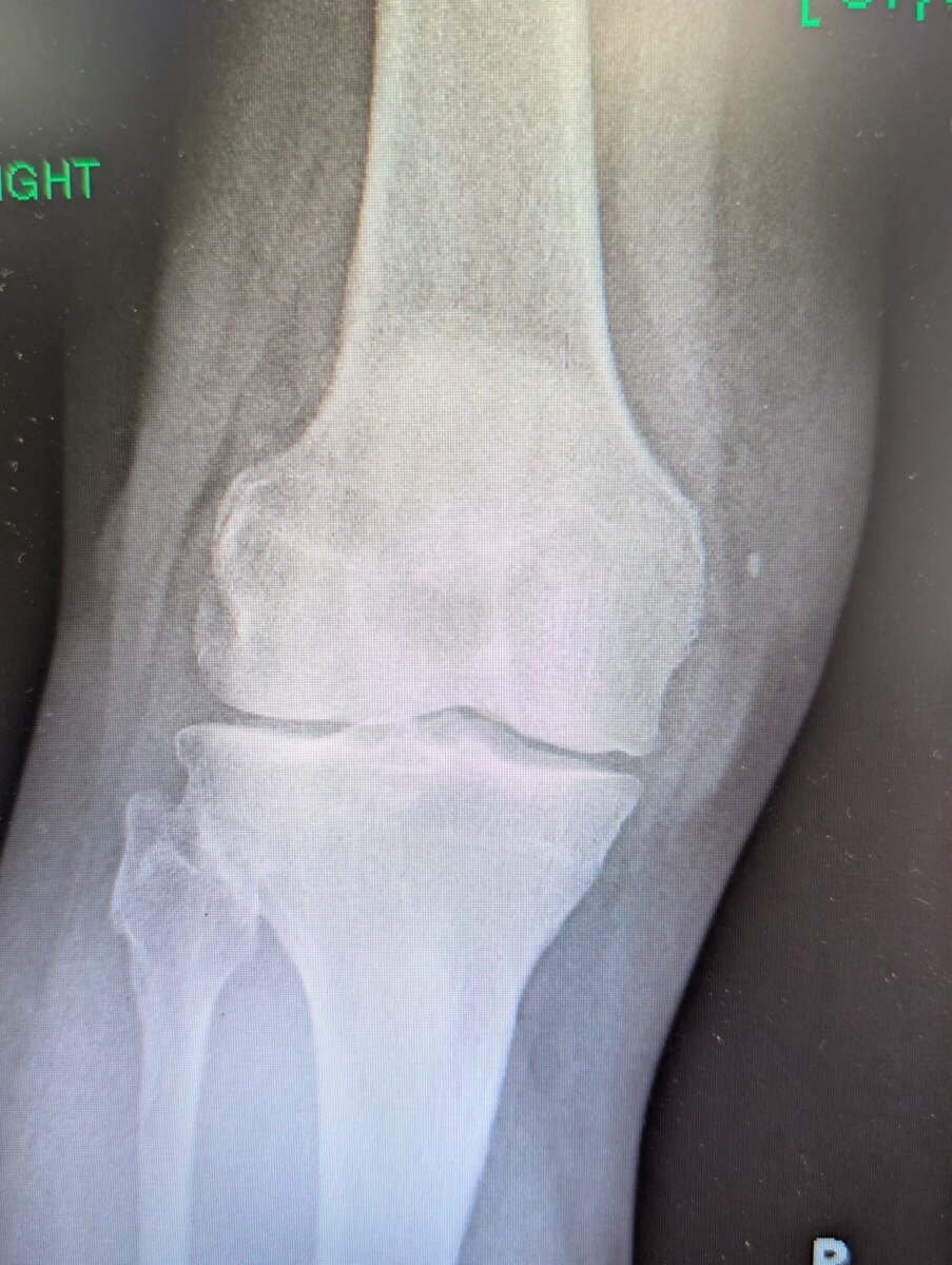
Weight-bearing radiograph of a knee with medial compartment arthritis
After
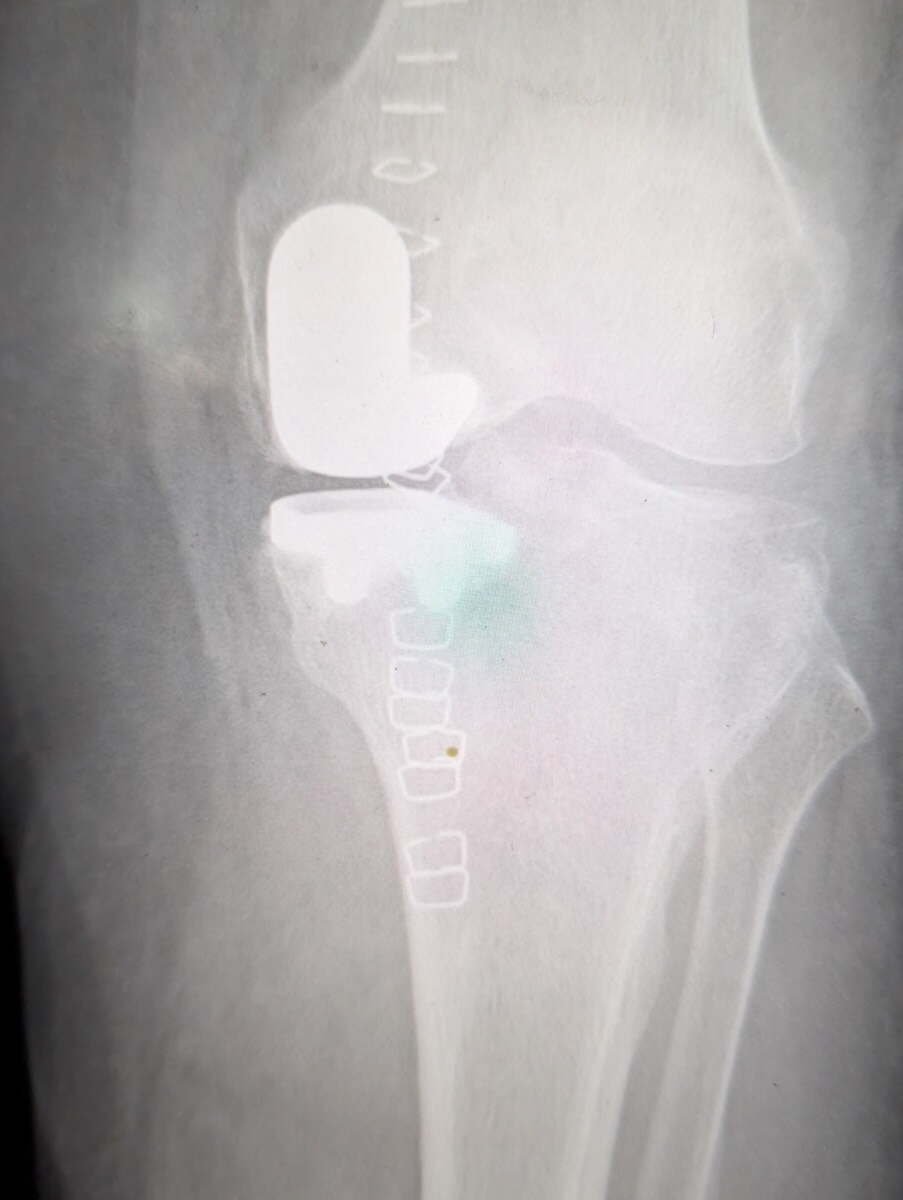
Radiograph of a robotic-assisted medial unicompartmental knee replacement
Partial Knee Replacement

Arthritis confined to the inner half of the knee. Only that compartment is resurfaced; the ligaments and the rest of the joint are left alone.

Before
Weight-bearing radiograph of an arthritic knee before replacement
After
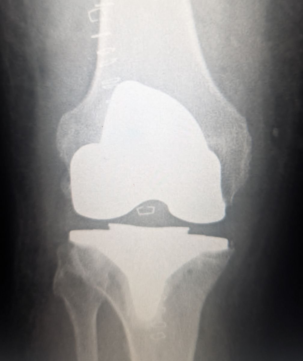
Radiograph of a robotic-assisted total knee replacement showing both femoral and tibial components
Robotic Total Knee Replacement

Arthritis across the whole joint. Planned in three dimensions and carried out with robotic assistance.

Before
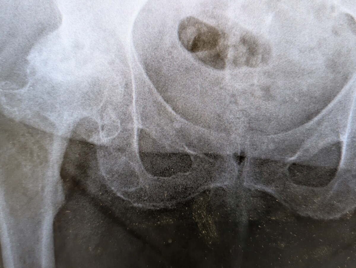
Radiograph of a hip with osteoarthritis and avascular necrosis
After
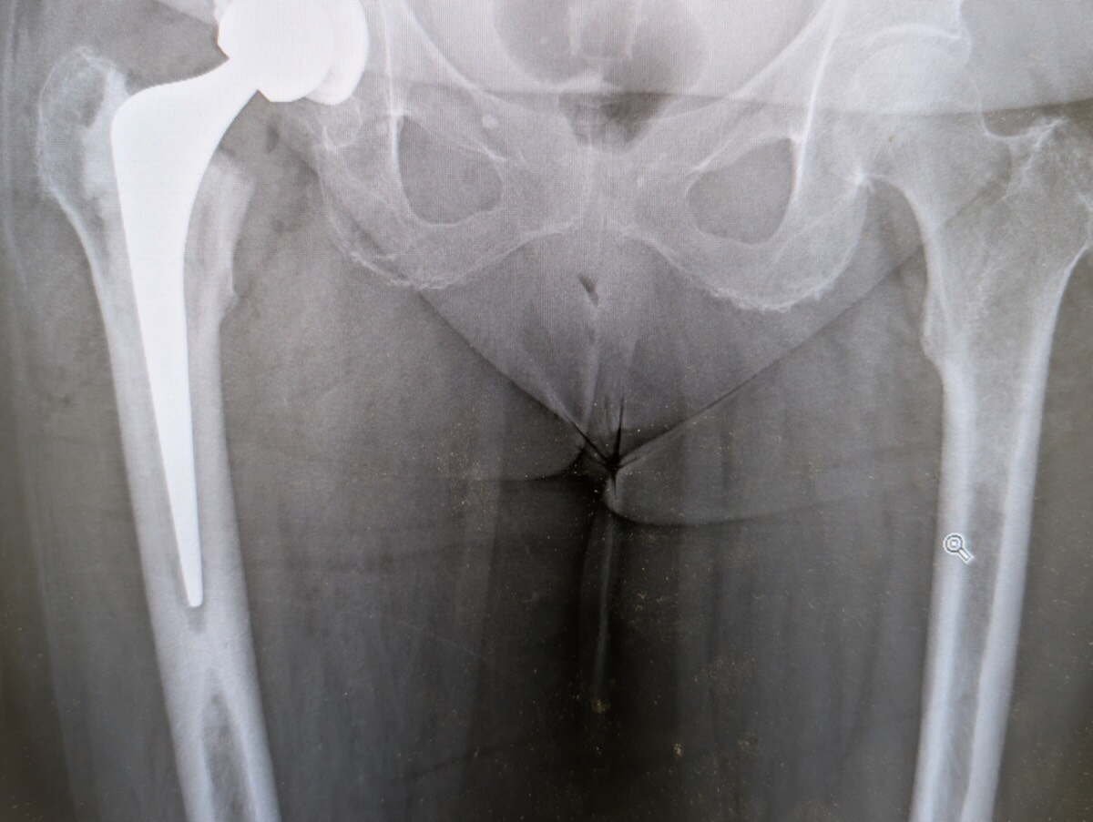
Radiograph of a robotic-assisted total hip replacement
Robotic Hip Replacement

Osteoarthritis with avascular necrosis of the femoral head, replaced with robotic assistance and close attention to leg length and offset.

Before
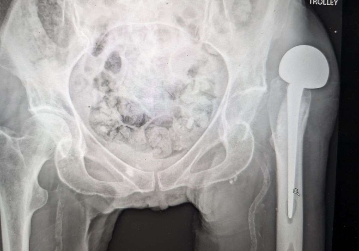
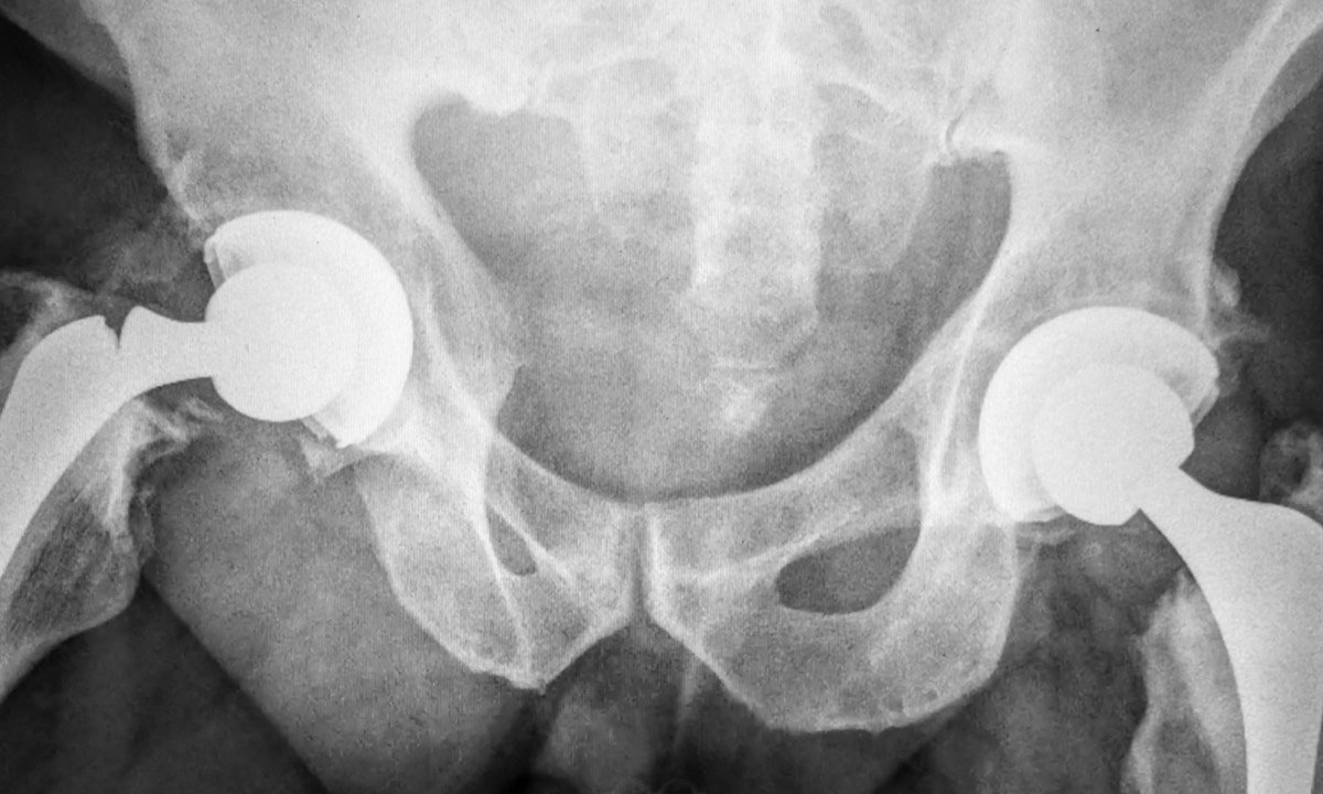
Radiograph of a dislocated hip hemiarthroplasty
After
Radiograph of a revision hip replacement using a constrained liner
Revision for a Dislocating Hip

A hemiarthroplasty that kept dislocating, revised to a total hip replacement with a constrained liner holding the head captive.

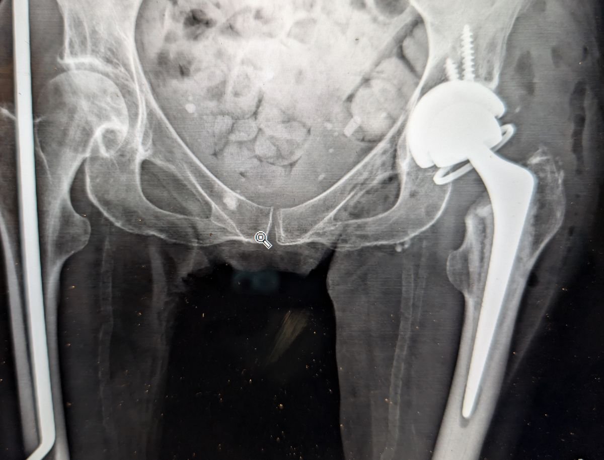
Radiograph showing a fractured Exeter femoral stem in situ
Fractured Femoral Stem

A cemented Exeter stem broken in the bone. An uncommon but demanding reason to revise a hip replacement.

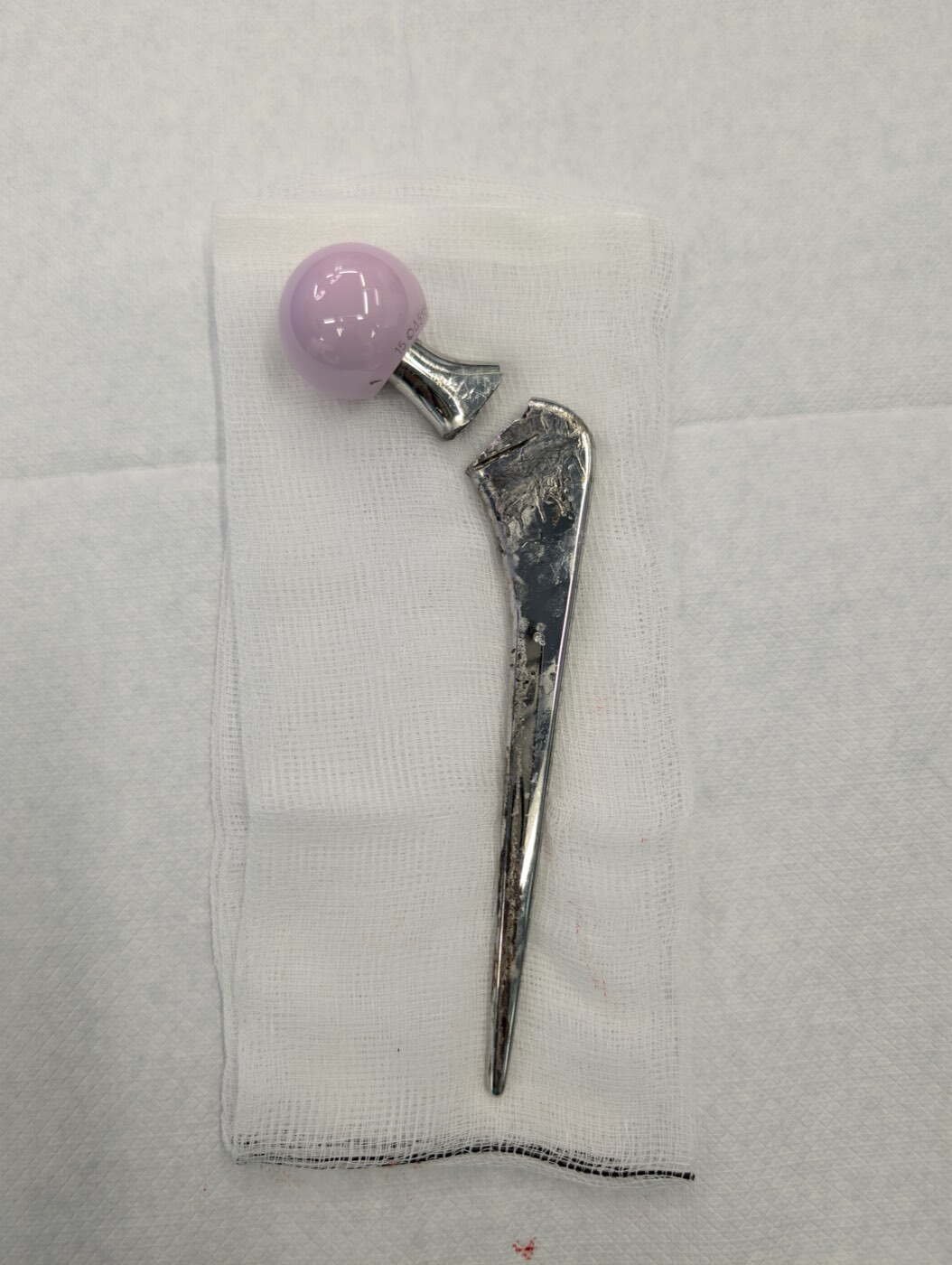
A fractured cemented Exeter femoral stem removed at revision hip surgery
The Stem, Once Removed

The same kind of fractured stem after extraction at revision surgery.

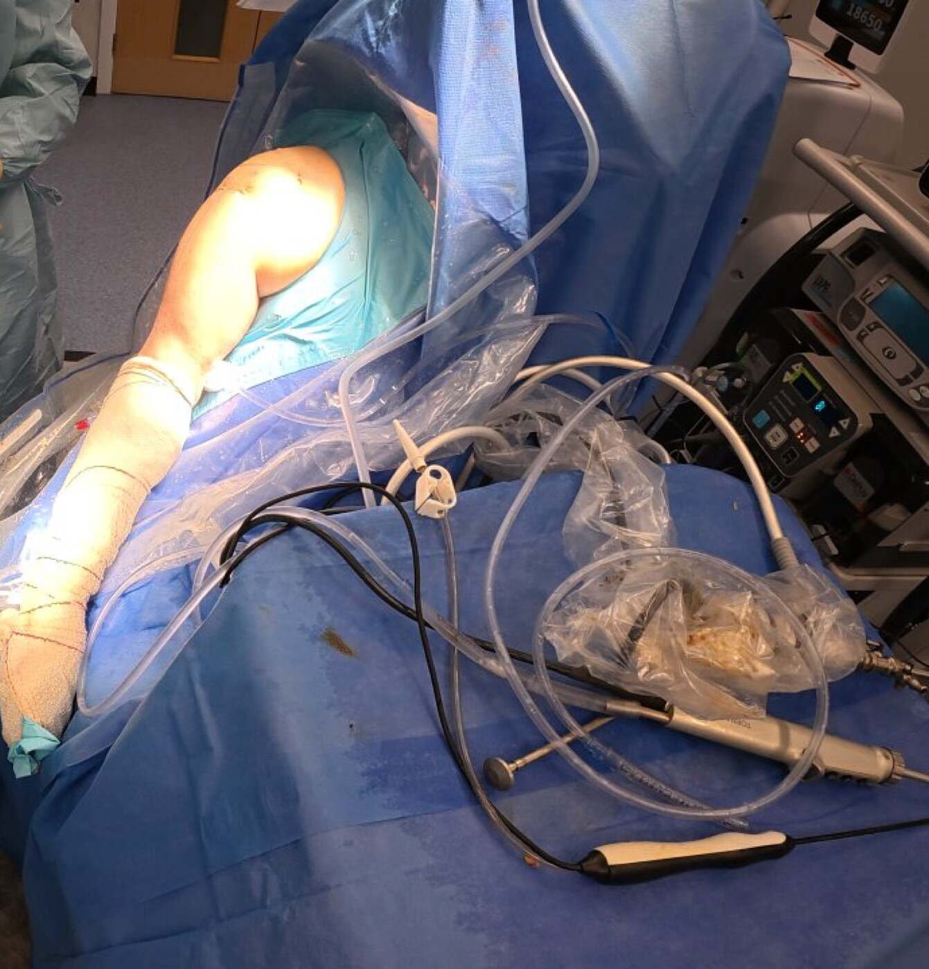
Operating theatre set up for shoulder arthroscopy in the beach chair position
In theatre

How the work actually looks

This is a shoulder arthroscopy set up in the beach chair position, with the patient semi-upright, the arm free to be moved through a full range, and the joint visualised on a screen through small portals.

Images on this page are deliberately restrained. Graphic operative photography has its place in surgical education, but it rarely helps a patient trying to decide whether to have an operation.

A note on these images. All radiographs have been cropped to remove any identifying information, and no patient is identifiable from anything shown here. Images are provided for general education only and do not represent a guaranteed outcome. Results vary between individuals.
Consultations across Delhi NCR

Bring your own scans to a consultation

If you already have X-rays, an MRI or a CT, bring them. A specialist reading of imaging you already own is often the fastest route to a clear answer.

Scroll to Top